
Lower Extremity Wound Management and Limb Loss
Abstract
Patients with multiple co-morbidities including Peripheral Arterial Disease, Diabetes, End-Stage Renal Disease, Dementia and poor or no mobility who develop lower extremity infections are at high risk for limb loss, which increases mortality. Conversion from BKA to AKA is common in these patients. In some populations AKA may be the preferred initial surgery, thus avoiding multiple surgical interventions with persistent failure to heal. However, no intervention reduces mortality other than preventing limb infection and wounds from occurring.
Introduction
Case Study 1: LW
LW is a 40-year-old. Female with DM, PAD, ESRD and previous R BKA. She developed necrotizing fasciitis of her left foot, which led to deep debridement and skin grafting, as well as balloon angioplasty. Her skin graft eventually failed, and she required L BKA. After two weeks her L BKA began to dehisce. AKA was discussed but the patient refused. Management of the dehiscence included debridement and betadine.
Case Study 2: DP
DP is a 66-year-old. female with PAD and Hepatitis C. A bypass graft of her left femoral artery became infected, and the infection spread to her foot. The foot wound was debrided and treated with a wound vac, which was ineffective. The patient underwent a BKA procedure, which had to be revised once due to poor healing and dehiscence.
Unfortunately, this is an all-too-common narrative in patients with multiple morbidities who develop lower limb infections. The aim of this paper was to review the risk factors and management options for lower limb infections and amputations; to assess the morbidity and mortality associated with limb amputation; to determine the incidence of BKA to AKA conversion; and to evaluate the necessity of BKA in patients for whom an initial AKA might be a more reasonable option.
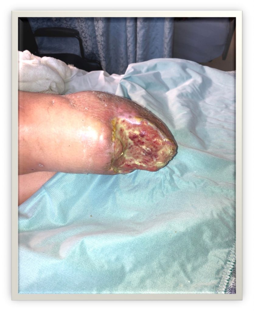
Image 1: Patient LW two weeks after dehiscence of left BKA
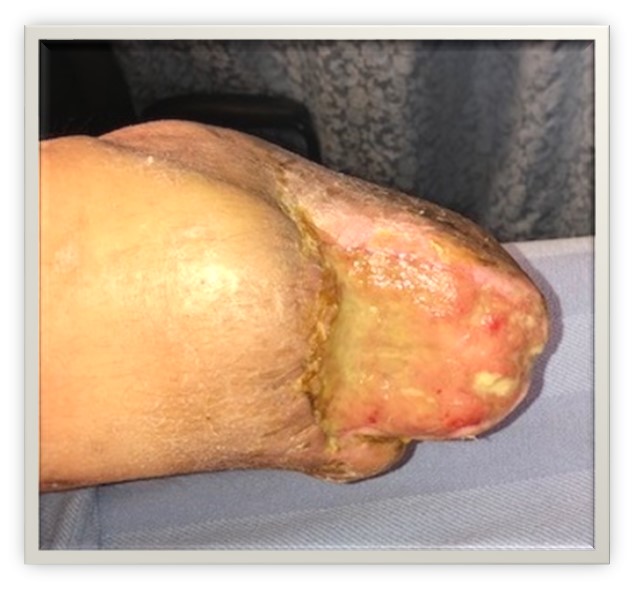
Image 2: Patient LW two months after dehiscence of left BKA. Managed with debridement of slough, betadine and calcium alginate.
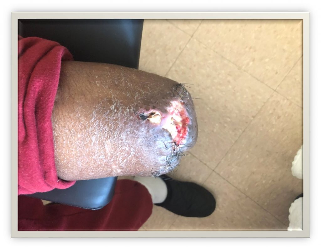
Image 3: Patient DP one week after BKA. Wound has dehisced and developed infection.
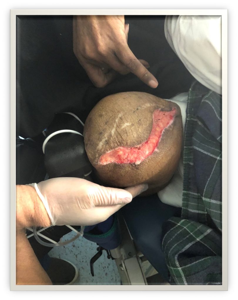
Image 4: Patient DP two weeks after BKA revision. Wound dehiscence has occurred again but without infection. Wound bed appears healthy but ultimately became infected, requiring an AKA.
Methodology
Data on the two case studies was collected, including the images shown here. An internet academic literature search was performed with the following keywords: BKA, AKA, BKA to AKA conversion, lower extremity wounds, lower extremity limb loss, management of lower extremity wounds. Studies that focused on patients with DM, PAD, ESRD, Dementia and/or Long Term Care Facility residency were included in the analysis, while studies of lower limb loss due to trauma, combat or for other reasons were excluded. A total of eleven (11) articles were selected. Information in the articles was organized into the following outline: Common Progression of Limb Loss; Incidence of Amputation; Primacy of BKA; Palliative Nature of Lower Limb Amputation; BKA to AKA Conversion; AKA Instead of BKA; Outcomes of the Cases.
Results
Dementia and residence in long-term care facilities often develop foot or limb infections that result in progressive amputation. Multiple interventions including wound care, debridement, angioplasty and/or revascularization, and wound bed treatments such as Integra and wound vacs often fail, necessitating amputation.
Estimates of annual lower limb amputations in the US range from 60,000 to 185,000. Once a foot is lost through BKA, preservation of the lower stump is desirable to facilitate rehabilitation and/or prosthesis placement, as well as to mitigate psychological trauma of AKAs, which generally have a higher mortality rate than BKAs.
However lower extremity amputation of any kind is usually a palliative procedure. At least one study found that half of all patients with PAD died within one year of lower extremity amputation. Patients with DM and ESRD on dialysis have particularly dismal survival rates.
BKA to AKA conversion is roughly 20% in many studies. Conversion usually occurs because of infection. Necrosis can be managed with general wound care and does not necessarily predict eventual AKA.
One study found that an initial AKA may the preferred treatment in non-ambulatory patients with ESRD, obviating the high failure rate of BKAs in these patients, as well as their increased mortality from repeated operations.
Proceeding directly to AKA in some patients presents ethical and psychological challenges, yet may be more humane than “whittling away” at the limb of a seriously ill or debilitated patient. Consideration of “AKA first” may be a reasonable approach on a case-by-case basis with informed discussion with patients, caregivers and advocates.
Conclusions
- Patients with multiple morbidities including DM, PAD, poor mobility, dementia and ESRD who develop wounds of the feet are at a high risk for lower extremity amputation.
- Necrosis is not a guarantee of amputation, but infection often is.
- Once amputation begins, mortality increases markedly.
- Sicker patients will likely not mobilize after BKA and their surgical sites will deteriorate.
- In certain patients (ESRD, extensive tissue loss, no ambulation) proceeding directly to AKA may be the more humane option.
References
Nehler M, Coll J, Hiatt W, Regensteiner J, et al. Functional outcome in a contemporary series of major lower extremity amputations. Soc Vas Surgery. 2003.
Kristensen M, Holm G, Kirketerp-Moller K, Krasheninnikoff M, et al. Very low survival rates after non-traumatic lower limp amputation in a consecutive series: what to do? Interactive CardioVascular and Thoracic Surgery. 2012; 14: 543-547.
Schuyler Jones W, Manesh P, Dai D, Vemulapalli S, et al. High mortality risks after major lower extremity amputation in Medicare patients with peripheral artery disease. American Heart Journal. 2012; 165(5): 809-815
Keagy B, Schwartz J, Kotb M, Burnham S, et al. Lower extremity amputation: The control series. Journal of Vascular Surgery. 1986; 4(4): 321-326.
Schofeld C, Libby G, Brennan G, MacAlpine R, et al. Mortality and Hospitalization in Patients after Amputation. Diabetes Care. 2006; 29(10): 2252-2256
Aulivola B, Hile C, Hamden A, Malachi G, et al. Major lower extremity amputation. Arch Surg. 2004; 139:395-399.
Rosen N, Gigi R, Haim A, Salai M, Chechik O. Mortality and reoperations following lower limb amputations. IMAJ. 2014 Feb; 16: 83-87.
Vishwakarm N, Rehmani B, Ararwal A, Ray JP. Reamputation rates, morbidity and rehabilitation after lower limp amputations. Intl Surg J. 2019 Feb 28;6(4):1274-9.
Yip V, Teo N, Johnstone R, Robertson A, et al. An Analysis of Risk Factors Associated with Failure of Below Knee Amputations. World J Surgery. 2006; 30:1081-1087.
Wu, J, Wong M, Zhiwen J, Wei-En W, et al. A Series of 210 Peripheral Arterial Disease Below-Knee Amputations and Predictors for Subsequent Above-Knee Amputations. Ann Vasc Dis. 2017; 10(3): 217-222.
Ploeg AJ, Lardenoye JW, Vrancke P, Breslau PJ. Contemporary Series of Morbidity and Mortality after Lower Limb Amputation. Eur J Vasc Endovasc Surg. 2005; 29: 633-637.

Stay up to date on the latest in wound care.
Join our mailing list today!


Thank You For Successfully Registering!
Stay tuned for the latest news and treatments in wound care tailored for medical professionals like you.


